Endoscopic/Microscopic Stapedotomy – Surgical Steps
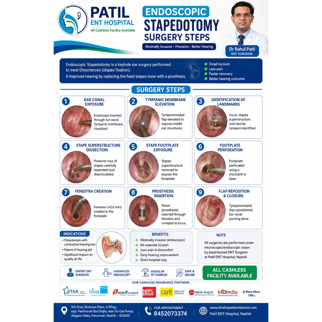
- Tympanomeatal flap elevation
- The ear canal is exposed and the tympanomeatal flap is elevated to access the middle ear.
- Middle-ear exposure
- The ossicular chain is inspected. Adequate visualization of the stapes, oval window, facial nerve and surrounding anatomy is obtained.
- Confirmation of stapes fixation
- Mobility of the ossicular chain is assessed and fixation of the stapes consistent with otosclerosis is confirmed.
- Incudostapedial joint separation
- The joint between the incus and stapes is carefully separated.
- Stapedius tendon division
- The stapedius tendon is divided to allow controlled removal of the stapes superstructure.
- Stapes superstructure removal
- The stapes crura/superstructure are carefully divided and removed while protecting the footplate and nearby facial nerve. (NCBI)
- Footplate fenestration (stapedotomy)
- A small fenestra is created in the stapes footplate using an appropriate laser, perforator or microdrill technique. The exact size and location depend on the surgeon's technique and prosthesis. (NCBI)
- Prosthesis measurement and selection
- The required prosthesis length is measured according to the distance between the incus and footplate, and an appropriately sized piston is selected. (NCBI)
- Piston prosthesis placement
- The piston is gently positioned through the footplate fenestra and attached securely to the long process of the incus.
- Mobility check
- The reconstructed ossicular chain is carefully checked for appropriate movement and stable prosthesis positioning.
- Flap repositioning
- The tympanomeatal flap is returned to its anatomical position and the external auditory canal is appropriately dressed/packed. (NCBI)
PATIL ENT HOSPITAL
Stapedotomy for Otosclerosis
Restoring sound transmission through the middle ear
All Cashless Facility Available
📍 5th Floor, Business Plaza, A Wing, Opp. Panchavati Bus Depot, Near Go Gas Pump, Adgaon Naka, Panchvati, Nashik – 422003, Maharashtra
Keywords